The most basic form of microscope is the hand lens. I use a ×10 lens; they are also commonly available in ×5 and ×20. I have found if I am careful I can actually get somewhat usable photographs by holding my hand lens between my phone camera and the specimen, and playing around with angles and distances. This requires holding my phone in one hand, and both the lens and the specimen in the other — a bit difficult, but it allows a lot of control over angle and lighting. Alternatively, if I can still get a decent angle, I set the specimen on my knee and just hold the lens. These are the techniques I use in the field and it can be enough to photograph the conidiophores of Peronospora.
For more detail and magnification you will want to use a proper microscope setup. However, you may not always want to mount a specimen on a slide. You can often get decent images by simply placing plant tissue under the microscope and having a look. This would be easiest with a stereo microscope, but I have found it works okay with my optical transmission microscope (i.e. a microscope designed for slides). This technique is particularly useful when you cannot see what part of the leaf to mount on a slide, when the pathogen structures are at a low density across a wide area. Obviously it works best when these structures are relatively large.
This is when you take several images focused at different depths and combine them using software to create a new image that has a larger depth of field. This can be particularly useful for microscopy without slides as the depth of field can be very poor if you are using a microscope designed for slides, where everything is flattened by the coverslip. It can also be useful with slides. Focus-stacked images are not primary data and you should always clearly indicate that you have used this method, especially in scientific publications. There is a useful blogpost on focus stacking here.
The way I normally make slides for rusts and powdery and downy mildews is to hold the infected part of the leaf over a glass slide and gently scrape the spores onto the slide with a razor blade. It can be difficult to avoid cutting into the leaf itself so this takes some practice. I then drop a small amount of water onto the spores on the slide and add the cover slip on top.
Often multiple different stages of rusts will be growing on the same leaf, visible as slightly different colours. It can be useful to isolate these from each other and look at each in detail. Chris Preston suggests using very fine tweezers to pick spores from a particular part of the leaf, and drop them onto a drop of water on a glass slide. I have also used tweezers to pick individual downy mildew conidiophores from a leaf, which can be useful when they are very few, although it can damage them quite severely.
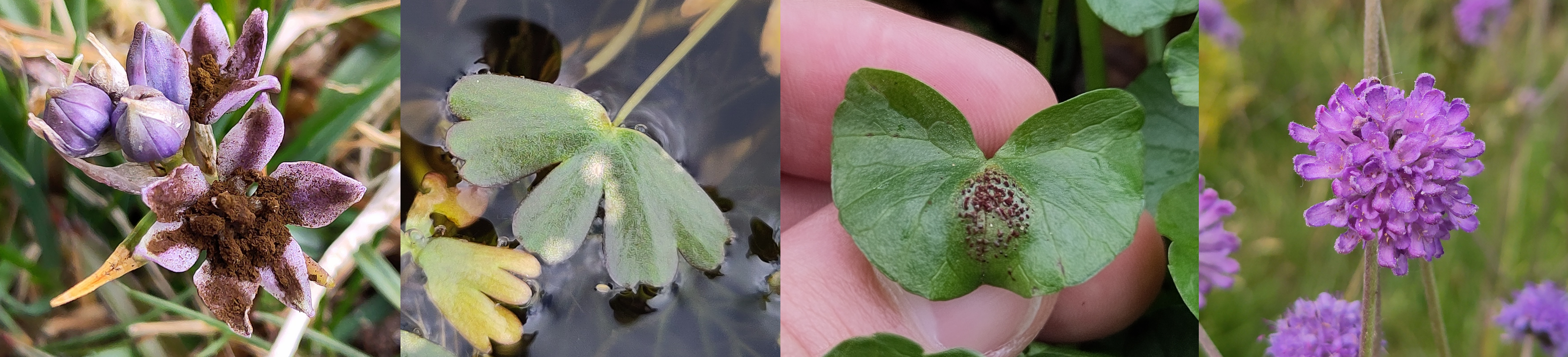
Some groups, like Ramularia, have minuscule spores produced on the outside of the leaf that can be difficult to transfer onto a slide. One trick that people use is to gently press a piece of sticky tape onto the leaf and then look at it under the microscope. The tape picks up the spores and sometimes their arrangement in clusters can be preserved.
The leaf spot fungi are particularly difficult to view under the microscope. Most of them produce spores tightly packed into small pycnidia embedded in the leaf tissue (left-hand figure). Pycnidia should be visible under the hand lens as small black or brown dots.
It is best to cut leaf spots with a razor blade or scalpel while they are still dry: this prevents the cut bits sticking together and allows you to make extremely thin sections. Hopefully some of these will slice through pycnidia. You can then add water and place a coverslip on top. The spores of Septoria and most other leaf spots are small, very thin, and colourless (right-hand figure), so you should be methodical and patient when looking for them.
Sometimes despite your best efforts you will find all of the pycnidia on the slide are intact, or the conidia are trapped inside the ones that have been sectioned. To fix this, place the slide on a flat surface and using the blunt end of a pencil or something similar, deliver a sharp but gentle tap to the coverslip. Be careful not to overdo this and break the coverslip! This method can break open pycnidia and force the spores out.
Often spores will fall out of a pycnidium to the bottom of the slide, so focusing the microscope on the bottom can make it easier to find them. Sometimes they will be more concentrated around an open pycnidium and following the trail can be a way to find pycnidia.
It can be difficult to see the septa in the conidia of some species of fungi. This is an important feature for identification so this can be an issue. Outer Hebrides Fungi has an example of conidia of Septoria arundinacea in Lugol's solution here, showing how it can be used to make the septa very clear and easy to count.
Entyloma produces its teliospores throughout the affected leaf tissue. I have found the best way to look at these is to again chop a small piece of tissue up, but then to place the cover slip on top and “smear” it by pressing firmly and evenly across the coverslip and moving it gently up and down with respect to the slide.
SEM is different to the other types of microscopy on this page in that it does not use light. Instead, a beam of electrons is fired at the sample and the resulting signals are used to understand the shape of the sample and some aspects of its composition. In order for this to work the sample is first coated with a very thin layer of a metal, often gold. Obviously, this is relatively expensive: at my university, the cost to the department for a single run of four samples is at least £20. The scope I have used in my department is a JSM-IT200, which I can say is a joy to use and very beginner-friendly. It is very hard to find prices for these things online but I think an equivalent machine new would cost somewhere in the mid-tens of thousands of pounds and up, rather outside of the price range of any individual who hasn't won the lottery. They are really machines meant for large research organisations like universities. Furthermore, without a library of SEM images to compare to, it can be difficult to interpret them when all you can compare them with is light micrographs and written descriptions based on light micrographs.
On the other hand, SEM gives you a level of detail you will not even get close to with light microscopy. This could be very useful for rust ID if there was a reference library for all of the species known on a given host. Currently, this does not exist. In summary, I think it would be useful if people studying rusts made an effort to build this reference library, and I think anyone describing new rust species should make an effort to include SEM images of spores in their description (not unreasonable given it only takes a few hours and maybe 20 quid). That said, SEM is not something anyone just trying to ID something should bother with.