

There are so many host plants in grasslands that it would be impossible to include them all, so I have just included the easiest to recognise pathogens that I see often on common hosts.
Somewhat uncommon. This is apparently most common on cultivated Daisies, but is found on wild plants too.
Extremely common, including in gardens and parks. Rather variable; look for warty, distorted leaves with orange or brown lumps.
This floricolous downy mildew is the most spectacular of all the plant pathogens I have seen. It perfectly and fundamentally changes its host's floral morphology, causing the petals to become longer and pinker, and the anthers to shrivel and abort. The result looks like a different plant altogether, a warm purple hemisphere of gently curving petals. This contrasts with the cooler purple petals fringed by a halo of magenta anthers in uninfected plants. It is widespread and common in large populations of the host but generally at a low prevalence. At the extremely large Succisa population at Murlough NNR it maybe infects somewhere around 0.5-1% of hosts.
Infection is systemic, so all flowerheads on the same plant are affected. The oömycete grows in the host xylem1, producing very little tissue until the host flowers. The infection is long term; Horáková and Skalický observed that the same individuals in a population of the host were infected each year over a three year period1.
This is my favourite plant pathogen, so it deserves an appropriately flattering name. I think Crown-of-amethyst describes its appearance well, in a way that contrasts with uninfected plants.
The density of the conidiophores produced on the petals varies quite a lot, but with a hand lens and careful searching you should be able to find them. Under the microscope the oöspores are clearly visible as purplish brown spheres embedded in the petals tissue. They are produced at a high density, and presumably rest in the soil1.
Somewhat common in large populations of the host, and very obvious when it is present. This smut produces its white powdery spores in the host's anthers, giving the appearance of a white halo around the flowerhead.
There is actually another flower smut on this host: Microbotryum flosculorum. It causes the petals to curl inwards and the flower to not open properly, and produces brown spores in the anthers.
Extremely common wherever the host is found. Look for dark brown, powdery anthers and dirty petals; in uninfected plants the anthers are yellow to deep orange. This species also apparently infects other members of the Caryophyllaceae — for a full list of hosts see the Plant Parasites of Europe entry. I have only found it on Stellaria graminea.
Look for pale, slightly downturned leaves on White Clover Trifolium repens. The conidiophores are whitish and can form on the top or underside of the leaflets. Rare wherever the host is found, but certainly also underrecorded as the symptoms are subtle.
Nearly ubiquitous in Ireland. Look for black spots on the leaves, which under the hand lens have small white synnemata. You may need to check quite a few black spots to find synnemata.
Fairly common but can be hard to spot. Whitish to pale beige conidiophores, sometimes on yellowed spots.
Common and obvious on Greater Bird's-foot Trefoil L. pedunculatus. Look for yellowed or dead patches on the upper surface of leaves, which have very dense white conidia underneath.
On both Greater Bird's-foot Trefoil L. pedunculatus and Common Bird's-foot Trefoil L. corniculatus. Look for yellowed, downturned leaves with purplish conidiophores underneath.
This was very common in 2024, but I don't remember seeing it before. I suspect it might vary in abundance a lot from year to year. Look for orange pustules on the leaves, bracts, and stems of the hosts.
Reasonably common where the hosts are found. Look for slightly yellowed, downturned leaves and a white down underneath, which is sometimes vein-delimited (left). I have only found P. euphrasiae on grassland Eyebrights, not on heathland species. Of the taxa found on Orobanchaceae, as far as I know only P. euphrasiae on Euphrasia spp. is distinguished from P. densa sensu stricto, and the rest are recorded as P. densa pending genetic work to determine which are distinct species.
This is a distinctive species that forms a black network of fungal tissue in the veins of the host leaves. It seems to be pretty common, particularly in the late Autumn and Winter.


Despite being so distinctive, this doesn't have an English name yet — let's call it Plantain Black Vein Fungus, calquing the German name here.
Brown leaf spots with white conidia in clusters. I have also found it growing on the flower stem.
A ubiquitous complex on most species in this host genus. They cannot be separated without microscopy.
There are a lot of different species infecting Buttercups. To avoid confusion I have given the three groups of smuts English names that I think best describe their appearance. Take your time with Entyloma in particular, the situation is pretty complex but most specimens are identifiable without microscopy. The one smut I have left out here is Entyloma verruculosum which I have not found yet; it causes slightly yellowed to dead brown, vein-delimited leaf spots and has warty spores2.
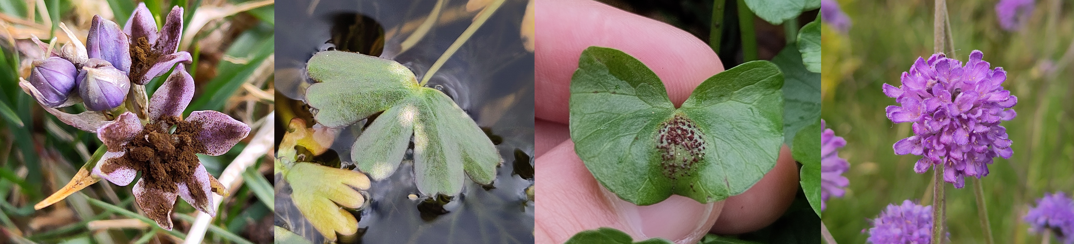
Swollen blisters which burst open to release brownish black spores. When held to the light, dark spores are visible inside the unopened blisters, distinguishing underdeveloped specimens from Wart Smuts Entyloma microsporum agg. Very common on Creeping Buttercup R. repens but quite rare on Meadow Buttercup R. acris.
Swollen “warts” which do not contain dark spores, distinguishing them from Blister Smut. Extremely common on Creeping Buttercup Ranunculus repens and apparently also found on Meadow Buttercup R. acris2.
Note this complex consists of two species, Entyloma microsporum and E. piepenbringiae, but the paper describing these does not give any way to distinguish them2. For now they are recorded on iNaturalist as complex Entyloma microsporum.
Yellowed spots above, with white conidia visible below. E. eburneum is reasonably common on Creeping Buttercup R. repens.
These are host specific, with each infecting a different species of Ranunculus. Note these were formerly all known as “Entyloma ranunculi-repentis” but this name is no longer valid.
A typical powdery mildew, with white mycelium growing over the leaf surface. The mycelium is often quite sparse and can be difficult to spot. Very common.
Look for yellowed, downturned leaves and dense beige conidiophores underneath. These can be distinguished by host. P. hiemalis is reasonably common on Meadow Buttercup Ranunculus acris, but P. ranunculi sensu stricto on Creeping Buttercup R. repens appears to be rather uncommon in my area at least. FRDBI records would suggest they are equally common.
Look for sad, lightly speckled and yellowed leaves. Brown apothecia are visible on the underside. Common on both R. repens and R. acris.
Very common. Bright orange aecia and uredinia, often following the veins of the leaf. This species also forms brown telia which are less easily found.


